Home
Common Techniques
Classroom Experiments
Virtual Experiments
Tutorials
Games
Glossary
Links
Publishing
Opportunities
About This Site
Contact Us
ZFIN
Cite Us
casanova Mutations in Zebrafish
Open protocol in HTML
Download the pre-lab worksheet
Background Information
In order to understand the nature of the casanova genes and mutations, one must first comprehend the nature of genetic mutations. In biology, mutations are changes to the nucleotide sequence of the genetic material of an organism. If this mutation occurs in a region of the DNA that contains information on how to make a protein, then the mutation can have an effect on the organism. In multi-cellular organisms, mutations can be subdivided into germ line mutations, which can be passed on to descendants, and somatic mutations, which are not transmitted to descendants. The mutations studied in zebrafish are typically passed down from parent to offspring, so they are germ line mutations.
The casanova gene is important to the proper development of the zebrafish cardiovascular system. cas mutants lack endoderm, which is necessary for proper migration of the heart precursors to the midline and for formation of the actual heart. Due to this deficiency, cas mutants heart precursors do not migrate, and cas mutants develop cardia bifida; the formation of bilateral hearts. Due to the severity of this condition zebrafish larvae with this disorder die relatively quickly. As the cas mutation leads the death of the zebrafish larvae, it is known as a lethal mutation. In addition, it is also referred to as a loss-of-function mutation because it causes the gene and the protein encoded by the gene to lose their functions.
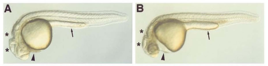
A: wildtype embryo at 24 hours post-fertilization
B: casanova embryo at 24 hours post-fertilizationPicture extracted from Alexander et al, 1999
The easiest way to identify casanova mutants is by the presence of pericardial edema, a clear mass posterior to the head and anterior to the yolk sac, which is not present in wild type zebrafish. Pericardial edema can be seen using bright field microscopy. The arrowhead in A points to the normal heart, and the arrowhead in diagram B points to the pericardial edema. The thin arrows point to the hindyolk, which is thinner in casanova embryos. The asterisks mark the ventricals of the brain, which form abnormally in casanova mutants.
Due to the advanced nature of this experiment, it is recommended that students read additional articles to gain a more comprehensive understanding of the subject matter.
Suggested Readings:
- Alexander, J., Rothenburg, M., Henry, G., and Stainier, D.Y.R. (1999). casanova Plays an Early and Essential Role in Endoderm Formation in Zebrafish. Developmental Biology 215, 343-357
- Glickman, N.S., Yelon, D. (2002). Cadiac development in zebrafish: coordination of form and function. Cell & Developmental Biology 13, 507-513.
Instructor Information
This experiment is suited for students who have a general knowledge of zebrafish development, including staging and organogenesis (specifically cardiogenesis). However, the expertise needed, both in knowledge and in laboratory skill, is minimal. This lab mainly tests microscopy skills and critical thinking during the analysis of the data. Therefore, this experiment would be most suitable for use in introductory developmental biology courses. Students may work in groups or independently depending on the materials available. Any class size may use this lab due to its small demand of materials.
Materials needed
- casanova mutant zebrafish embryos crossed into the actin:GFP transgenic line
- Compound Fluorescent microscope
- Embryo Loops and materials for making embryo loops (capillary tubes, fishing line and superglue)
- Fine Forceps
- Fish Water
- Glass slides
- Methylcellulose for mounting the embryos
- Timers or Stopwatches
- Stereomicroscopes
Techniques Used
- Creating embryo loops
- Introduction to compound microscope
- Mounting embryos in methylcellulose
- Dechorionation